
Product Design · Medical Device
CathNav
A multi-component surgical navigation system bridging digital planning, physical hardware, and real-time mobile guidance — designed across a three-month sprint to evaluate a novel approach to bedside EVD placement and support a patentability assessment.

The Challenge
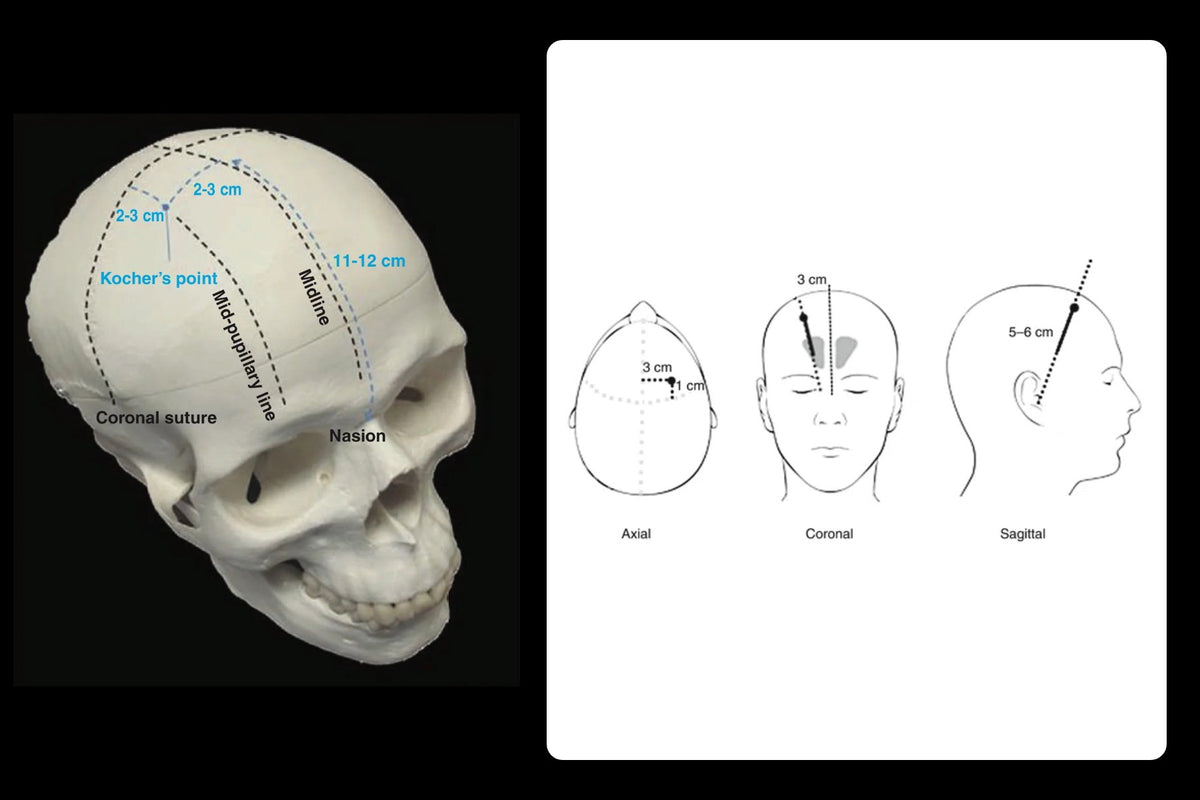
External ventricular drain placement demands precise targeting within the brain — errors can damage healthy tissue and cause serious, irreversible complications. Existing bedside methods relied on anatomical landmarks such as Kocher's point and surgeon experience, with minimal image guidance. The opportunity was to design a system that made CT imaging clinically actionable at the bedside — and to evaluate whether that system had sufficient novelty to support a patentability assessment and early-stage investor conversations.
Source: EVD complications review (NIH / PMC)Hemorrhage and misplacement
Catheter insertion carries a 10–40% risk of hemorrhage or misplacement along the tract. Rapid overdrainage can also cause a subdural hematoma.
Infection
Meningitis and ventriculitis risk ranges from 0% to 22%, roughly 8–10 per 1,000 drainage days, and rises with longer drainage (especially past 7 days), catheter changes and system breaks.
Blockage
Debris, blood clots or kinked tubing can stop CSF drainage and spike intracranial pressure.
Drainage level and transport
The drain must stay level with the tragus. Bed position changes and transport lapses, like an unclamped drain or lost head-of-bed elevation, cause over- or under-drainage and accidental dislodgement.

Storyboard
One EVD placement, from guesswork to guided, across the five phases CathNav was designed around.
-
The problem
Placed by feel
Bedside EVD placement relied on anatomical landmarks and surgeon experience, with minimal image guidance. A missed target can damage healthy brain tissue.
-
Phase 01
Plan on the CT
The surgeon imports the patient's CT, measures skull thickness, selects the catheter path and compensates for midline shift.
-
Phase 02
Register with the Smart Mat
At the bedside, the Smart Mat registers the patient's position so the plan can be mapped onto their real anatomy.
-
Phase 03
Fuse the scan to the patient
CT-to-anatomy spatial fusion lines the scan up with the patient's head, so the planned path matches where the patient actually is.
-
Phase 04
Position the guide
The sterile cath guide is set to the planned entry point and trajectory before anything is inserted.
-
Phase 05
Navigate live
A phone mounted at the bedside shows the catheter's progress against the planned path in real time during placement.
System Design & User Flow
Designed a five-phase clinical user flow connecting digital and physical touchpoints: CT-based digital procedure planning, Smart Mat patient registration at the bedside, CT-to-anatomy spatial fusion, sterile cath guide positioning, and live catheter navigation on a mounted mobile device. Each phase was designed to unlock sequentially — maintaining clinical focus and reducing cognitive load during a high-stakes procedure.
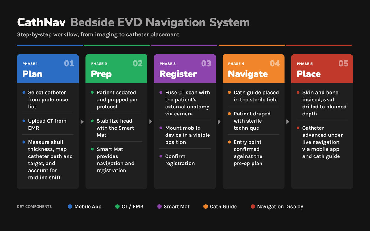
Prototype Design & Wireframes
Wireframed and prototyped the system's mobile-first interaction model across the full procedure — CT import and digital planning (skull thickness measurement, catheter path selection, midline shift compensation), Smart Mat-based spatial registration, and live mobile navigation during placement. Every interface decision balanced clinical precision with usability under sterile, high-pressure conditions. Hardware and software touchpoints were designed together to evaluate the full system before any manufacturing investment.


Cath Guide: Form & Interaction
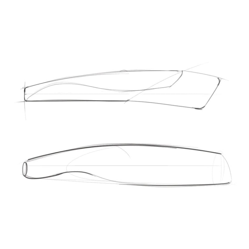
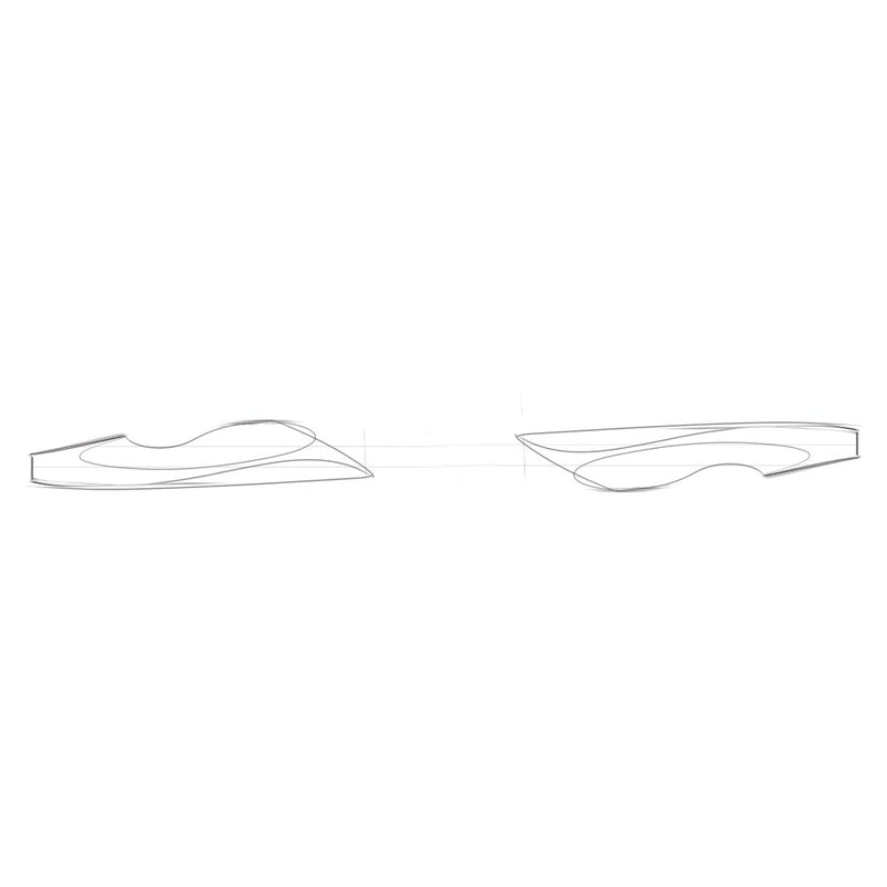
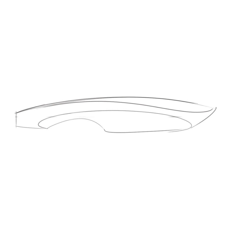
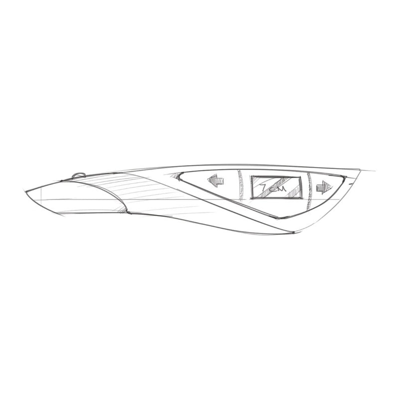
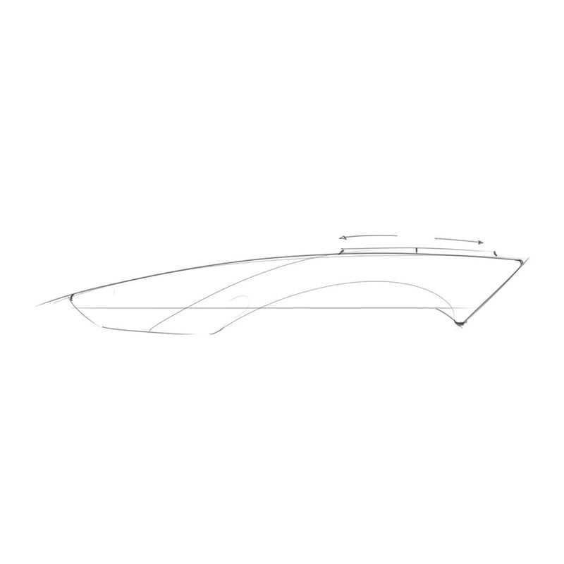
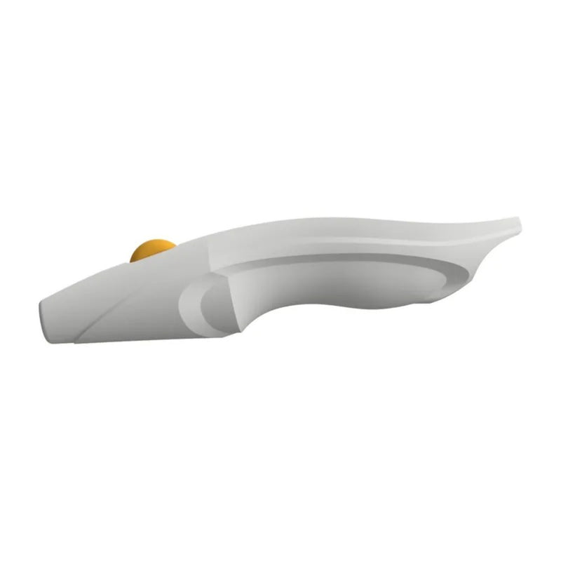
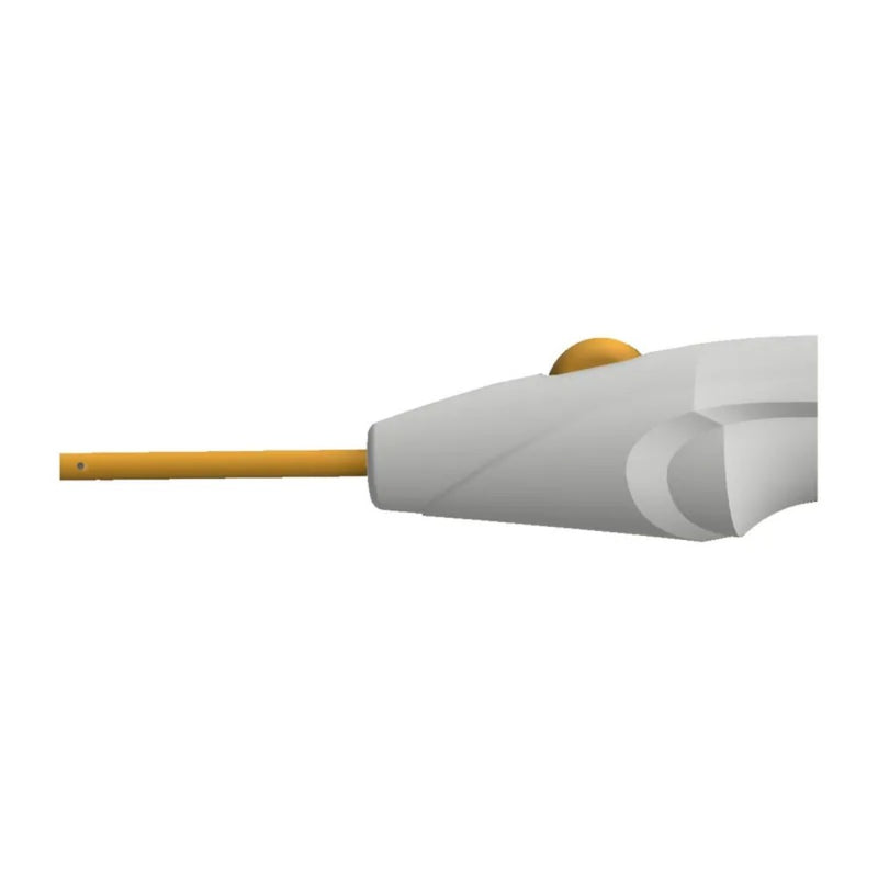
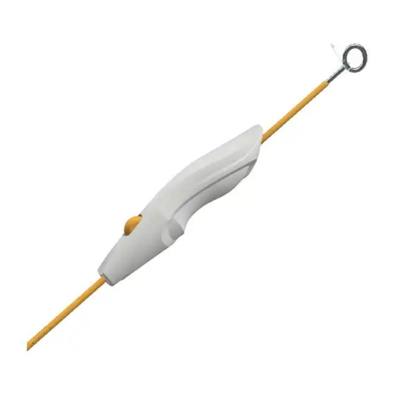
The handheld cath guide holds the catheter on the planned path while the phone tracks it. Sketches explored a pen grip that sits low in the hand for fine control at the entry point, then 3D models resolved the index-finger wheel and the catheter path through the body.
Pen grip
A slim, sculpted body held like a pen, so the hand rests close to the entry point for fine angle control.
Index-finger wheel
A wheel set into the body, like a mouse wheel, advances the catheter in small, controlled steps.
Depth on the device
Early concepts put a small depth readout between advance and retract arrows, right where the eye already is.
Straight-through path
The catheter and stylet pass straight through the guide, keeping the tip on the planned trajectory.
Outcome
A fully articulated proof of system covering hardware, software, and procedural user flow, with the technical depth needed to assess the novel contributions of Smart Mat registration, CT-to-anatomy spatial fusion, and integrated cath guide navigation. It formed the basis for a formal patentability evaluation and early-stage investor conversations. Here is that system redesigned for 2026 as a working prototype: one guided flow, a light, high-contrast UI built for the bedside with true-to-life CT imaging, and actions that stay locked until it's safe to continue. Click through a full placement, from case setup to marking the catheter placed.
- Plan
- Register
- Fuse
- Guide
- Navigate
Set up the case
Confirm the patient, scan and guide, then finish bedside prep.
Bedside prep2/3 done
Find the Smart Mat
Hold the phone steady above the patient's head until all four markers lock.
Catheter placed
Placement is logged to the case with the plan, fusion and live track.
Try the prototype
- Set up the case
- Plan the path
- Register on the Smart Mat
- Check the fusion
- Position the guide
- Navigate to target
2026 redesign concept of the CathNav app. All values are illustrative.
- 1Landmarks only. Today, entry and angle are measured off the skull and judged by eye in three planes.
- 2Ventricular catheter. Marked tip gives a visual depth reference.
- 3Index-finger wheel. Set into the body like a mouse wheel to advance the catheter in fine steps.
- 4Stylet. Keeps the catheter rigid on the planned path, then pulls free.
- 5CathNav app. Ties it all together: loads the CT plan, registers the patient on the Smart Mat, pairs with the guide and tracks the catheter live.